
Contact

Did you know...
The average heart beats 72 times a minute; 100,000 times a day; 36 million times a year, 2.5 billion times during a life time.
Structural Disorders of the Heart
Structural disorders of the heart can affect people of any age. Congenital heart defects are present from birth whereas valve disorders tend to arise later in life.
Congenital Heart Defects
Some types of congenital heart defects are familial, suggesting a genetic link is very possible, yet a number have no obvious cause. There have been cases where the mother has passed on an infection during pregnancy or through exposure to certain drugs; including alcohol, which have both lead to birth defects. Some symptoms of CHD include shortness of breath which often leads to difficulty in feeding and a slowed weight gain.
Ventricular Septal Defect (VSD)
This defect involves a hole in the muscular wall between the two ventricles leading to a mixing of the blood. Oxygenated blood from the left ventricle flows through the hole, resulting in too much blood being pumped by the right ventricle into the lungs. The scale of the hole varies between individuals but a small one may close as the child develops but a larger one tends to require surgical repair.
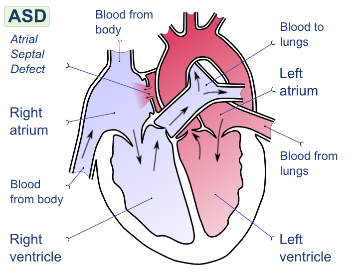
Atrial Septal Defect (ASD)
This is an abnormal opening in the wall between the two atria. There is an increased blood flow to the lungs as blood bypasses from the high-pressure left atrium to the right. This results in limited blood being transported around the rest of the body, and therefore minimal oxygen and nutrients reaching important organs and tissues. Both atrial and ventricular septal defects are common in Down Syndrome children.
Coarctation of the Aorta
A short section of the aorta becomes narrowed. It usually occurs at the point where the main arteries branch off for supply to the head and upper body which subsequently results in restricted blood flow to the lower body. In response, the heart works extra hard to try to compensate so blood pressure in the upper body is raised significantly. A patient suffering from this CHD may experience difficulty breathing, eating and usually has a pale appearance. Urgent corrective surgery is usually needed.
IMAGE: A: Coarctation (narrowing) of the Aorta, 1: Inferior Vena Cava, 2: Right Pulmonary Veins, 3: Right Pulmonary Artery, 4:Superior Vena Cava, 5: Left Pulmonary Artery, 6: Left Pulmonary Veins, 7: Right Ventricle, 8: Left Ventricle, 9: Main Pulmonary Artery, 10: Aorta
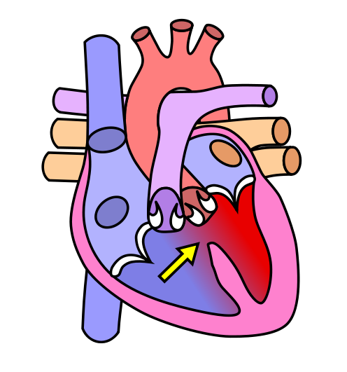
Tetralogy of Fallot
This CHD is a combination of four structural defects. It results in low oxygenation of blood due to mixing of oxygenated and deoxygenated blood in the left ventricle, caused by a ventricular septal defect and the desired pathway of flow through the aorta due to the obstruction to flow through the pulmonary valve (pulmonary stenosis). The other two defects include a thickened right ventricular wall and displacement of the aorta to the right, allowing deoxygenated blood from the right ventricle to flow into it. Only three of the four structural defects are always present in a patient with tetralogy of fallot, and symptoms include breathlessness and a distinct blue skin colour (cyanosis).
IMAGE:
A: Pulmonic Stenosis
B: Overriding Aorta
C: Ventricular Septal Defect (VSD)
D: Right Ventricular Hypertrophy
Valve Disorders
Valve malfunctioning is classed in two ways:
- Stenosis = the valve does not open properly and therefore restricts blood flow through a narrow passage. Often due to infection or can be congenital.
- Incompetence = or ‘leaky’ valve, refers to when the valve does not close properly, resulting in backflow of blood. As a result the heart has to work harder to circulate blood. Commonly occurs after a myocardial infarction or an infection of the valve.
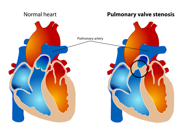
Image of Pulmonary Valve Stenosis by Mariana Ruiz LadyofHats [Public domain],via Wikimedia Commons
Conditions Affecting the Layers of the Heart
Inflammatory Heart Disease
This disease involves the inflammation of one of the layers of the heart wall.
Endocarditis- inflammation of the inner layer of the heart, causing most damage to the heart valves
Myocarditis- inflammation of the muscular, middle layer the myocardium
Pericarditis- inflammation of the pericardium; the protective sac around the heart
Inflammatory Cardiomealgy- general enlargement of the heart
VSD, Modified version by Dake of the original heart diagram by Wapcaplet via Wikimedia Common
By Manco Capac (Own work) [CC-BY-SA-3.0 (www.creativecommons.org/licenses/by-sa/3.0)], via Wikimedia Commons

Image from Wikimedia Commons

By Wapcaplet [CC-BY-SA-2.5-2.0-1.0 (www.creativecommons.org/licenses/by-sa/2.5-2.0-1.0)], via Wikimedia Commons